
Author: Qutub; Learner, Age 15 years
This is a self determined individual project chosen by the learner for independent research and documentation. The organization, questions and directions chosen are entirely those of the learner.
Note from author: The human body has blood which plays an integral role in sustaining life. We usually bleed when we get injured. I started bleeding when I got cut so I got curious to know more about blood. What is it? What is it made of and what does it do?. Also, how does the bleeding stop?. I want to learn more about blood because I am interested in biology.
My aims are : How is blood formed?. What is the blood made of, composition of blood?. How does the heart pump blood?. What does blood do in the body?. Where does blood flow?. What happens when foreign substances enter our body?.
Table of contents:
1: History of blood
2: Composition of blood
A: Plasma
B: Red blood cells
C: White blood cells
D: Platelets
3: The heart
4: Blood type
Blood
The study of blood is called hematology. The study of blood flow is called hemodynamics. A man has 12 pints (1 pint = 473 ml) of blood. A women has about 9 pints.The maximum amount of blood the human body can lose is 40% of the total amount. If it loses more than this, it won’t be able to function or remake that much blood. Therefore there is a high chance of fatality.
History of blood:
The first person to describe the blood flow in detail was William Harvey. He was an English physician who made lots of contributions in anatomy. Most physicians of the time thought that the blood was pumped by the lungs. Harvey, observing the motion of the heart in animals was able to see that the systole(contraction phase) was the phase of the heart pumping blood. Harvey’s predecessors had started observing the circulatory system but he was the first to describe it.
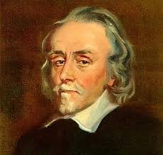
Source of image: http://md.rcm.upr.edu/romanfranco/wp-content/uploads/sites/41/2015/02/harveypic.jpg
Composition of Blood
Blood consists of 4 parts, Plasma, Red blood cells , White blood cells and Platelets.
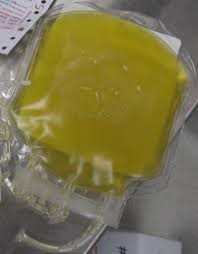
Plasma: Plasma contains water, proteins, glucose(sugar), hormones(chemical messengers) like insulin, carbon dioxide, oxygen, enzymes, solutes and electrolytes. Plasma also carries nutrients, gases and salts.
It’s the liquid part of blood and it’s pale yellow. It’s 55% of the blood in volume. It transports nutrients and helps to remove waste. It regulates fluids and electrolyte balance in the blood. (Electrolytes are minerals like sodium, calcium, potassium. You get them from the food you eat and what you drink. Electrolyte imbalance is caused by losing fluids like vomiting, loose stools or diarrhea and even increased sweating. Electrolyte balance is important because of hydration and muscle contraction etc. The muscles need electrolytes to function ).
The plasma maintains the pH of blood. pH is the abbreviation for potential hydrogen (concentration of the hydrogen ions). It’s the measure of the acidity or the alkalinity. The higher the pH the more alkaline and oxygen rich the blood is. The lower the pH the more acidic and oxygen deprived blood is. If the pH changes then this causes symptoms and diseases. The ideal pH of blood is 7.4. Neutral but a little more to the alkaline side.
Plasma proteins are the most abundant dissolved substances in plasma.Proteins make 7 to 9% of plasma. The proteins are albumin , globulin, and fibrinogen.
Albumin: The albumin helps to maintain osmotic pressure (minimum pressure which needs to be applied to a solution to prevent the inward flow of water across a semipermeable membrane (it allows certain molecules or ions to pass through it by diffusion ( A process in which molecules move from an area of high concentration to an area of low concentration) of blood. They are 60% of plasma proteins. They are produced in the liver. Albumin are the smallest but they are majority.
Globulin: Globulin are 36% of plasma proteins. There are 3 types- Alpha, beta and gamma globulins. Gamma globulin is produced by white blood cells. Gamma has an immune function. Gamma globulin is an antibody and they are Y shaped. The alpha and beta globulin are produced in the liver. Alpha and beta globulin transport lipids and vitamins. Lipids contain fat which is used by the body to store energy.
Fibrinogen: Fibrinogen is a soluble protein. If there is an injury to the blood vessel fibrinogen become fibrin and helping to form a blood clot. They turn into insoluble strands of fibrin. They are 4% of the total volume of proteins.
The solutes are: Sodium ions, bicarbonate, calcium ions.
The hormones like Insulin are chemical messengers (compound that transmits messages). If the body dehydrates then the plasma becomes concentrated so its osmolarity(total number of solute particles per litre.) will increase. The osmolarity increase is detected by osmoreceptors.The increased osmolarity results in thirst and release of an antidiuretic hormone [ADH] from the posterior pituitary gland. It is a water-regulating hormone made by the hypothalamus (in the brain), and stored in the posterior pituitary gland(master gland).
Enzymes: They are also called catalysts. There are many types of enzymes. Some that stop infections and some that break down food and others. Enzymes are proteins that speed up chemical reactions. Without them reactions would be too slow to support life. Cells also have enzymes in them. All enzymes have an active site. This is where substrates(material upon which an enzyme acts) join to them. There the substrates are broken down to form products. Each enzyme can only join with one substrate. Enzymes are made up of amino acids. Enzymes are made by stringing together 100 or 1000 amino acids in a specific and unique way. An example is creatine phosphokinase.
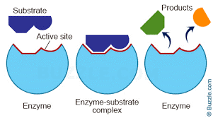
Source of images:
https://pmgbiology.files.wordpress.com/2014/12/enzyme-working-mechanism-e1417645715778.jpg
https://upload.wikimedia.org/wikipedia/commons/1/1d/FreshFrozenPlasma.JPG
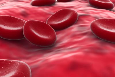
Red blood cells: The main function of the RBC is to transport oxygen throughout the body and to bring back carbon dioxide to the lungs. They are called erythrocytes. They are anucleated. Therefore they have more free space. This allows haemoglobin to be stored. Hemoglobin is a respiratory pigment (a molecule that increases the oxygen carrying capacity of blood). Hemoglobin binds to oxygen or carbon dioxide. This allows transport of oxygen throughout the body. This also allows the body to get rid of carbon dioxide.
Hemoglobin has iron in it, when binds with oxygen it gives blood its bright red color. RBC are one of the smallest cells in the body. Their size increases their speed. They are flexible so they can flow into small places and give oxygen everywhere. There is a disease called sickle cell anemia when RBC becomes rigid and it blocks the vessel. Each RBC has 280 million molecules of haemoglobin.
RBC are 6 to 8 micrometers in size. They are shaped like a biconcave disk. They have a depression in the middle. They are formed in the bone marrow by a process called erythropoiesis. They only last for 120 days because when they go into small places their outer membrane gets damaged and they tear apart.
These cells don’t have a nucleus so they can’t remake themselves. They do not have mitochondria (powerhouse of the cell). The RBCs functions are already programed so they don’t need a nucleus or a mitochondria. RBC are able to get energy to move around from fermentation(process that converts sugar to acids, They convert glucose to lactic acid). When the RBC is maturing it loses its nucleus and the mitochondria. Hemoglobin makes up 95% of the RBC cell.
In the lungs the oxygen goes into the cell by diffusion. The oxygen molecules help each other when one oxygen molecule binds to the haemoglobin and changes the shape of the haemoglobin making it easier for other oxygen molecules to bind. some oxygen molecules get dissolved into the plasma. This is how it’s transported throughout the body to muscles, tissues etc. It’s transported because the muscles and tissue need it for energy.
The muscles create carbon dioxide because of cellular respiration. This always happens even when you are asleep. Carbon dioxide particles compete with oxygen for the haemoglobin. They bind to it and oxygen falls off. This happens because the concentration of carbon dioxide is high. Then it goes back to the lungs. There the oxygen competes with the carbon dioxide for the haemoglobin. They bind to it and carbon dioxide falls off and gets exhaled. This happens because the concentration of oxygen is high. This is a continues cycle. The body needs to get rid of carbon dioxide because it is a waste product.

Source of image: http://www.daviddarling.info/images/red_blood_cells.jpg
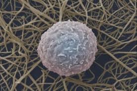
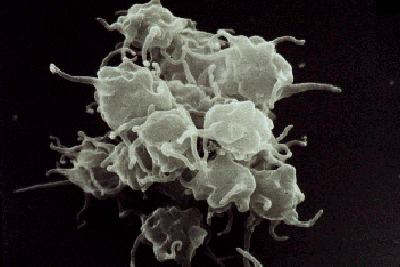
White blood cells: They are also called leukocytes. They are an important part of the immune system. They attack bacteria, viruses and germs. They are produced in the bone marrow. They have a nucleus so they can reproduce( mitosis) and they have DNA(deoxyribonucleic acid).
There are 6 main types of WBC that are distinct in form and function
- Neutrophils,
- Lymphocytes,
- Eosinophils,
- Monocytes,
- Phagocytes, and
- Basophils.
Neutrophils: They are 62% of the total WBC. Their main targets are bacteria and fungi. There life time is 6 hours to a few days. They kill by ingesting or by sending signals to B cells. They do this in the blood and the tissue. They act like surveillance cells looking for infections. They can sense a sight of infection because of the chemicals released by the bacteria.
Eosinophil: They are 2.3% of the total amount of WBC. Their main targets are larger parasites. They eat them or call B cells. There lifespan is 8 to 12 days.
Basophil: They are 0.4%. They release histamine when inflammation happens. They live for a few hours to a few days.
Monocytes: They are 5.3%. They migrate from the bloodstream to the tissues. They kill all pathogens in the tissue. Or they signal B cells. Their lifespan is hours to days.
Lymphocytes: There are 2 types of lymphocytes. T cells and B cells They are 30%. They live for 12 to 15 days. B cells create antibodies. Pathogens cause diseases. They release toxins. The antibodies then destroy them. T cells kill the cells that are infected not the pathogens.
When pathogens(bacteria, viruses or any microorganisms that can cause disease) enter the body, WBCs called phagocytes pass through the wall of the blood vessel into the tissue. There they ingest the pathogens.They also send signals to call other cells. Some cells that come are lymphocytes. They destroy pathogens sometimes directly.
Source of image: http://www.iflscience.com/sites/www.iflscience.com/files/styles/ifls_large/public/blog/%5Bnid%5D/5814247239_3a527eb991_o.jpg?itok=yFwBIMTF
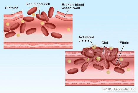
Platelets: Platelets are produced in the bone marrow by cells called megakaryocytes.They protect the blood vessels. They are fragments of a cell’s cytoplasm( the cell’s internal sub structure). They help in clotting. When your blood vessel gets cut then blood gets exposed to collagen, which is outside the vessel, which is the most abundant protein.
- When platelets come into contact with collagen they bind. The collagen has molecules that bind to platelets. They stick to each other. They also change shape. They may look like an octopus. This attracts fibrinogen.
- When they come to the site they become fibrin. They have sub units and they stick together to make a strand. It’s like fibrinogen except there is an extra piece in fibrinogen that stops subunits to stick together.
- They turn into fibrin on the attack site because of a chemical reaction that happens with tissue factor(key initiator)(Tissue factor is a protein present in the tissue, also called thromboplastin). The tissue factor call others who call others etc. They help in the chemical reaction. In the end the molecule that turns fibrinogen into fibrin is a protein called thrombin.
- The fibrin strands come and stick to the sticky platelets forming something like a mesh. When this happens red blood cells get trapped in therefore forming a blood clot. None of these fibrinogen stick together inside the blood because there is no tissue factor in the blood. There is also no collagen in the blood so the platelets don’t stick together in the blood either. A blood vessel is made up of endothelial cells. They are packed together. They have a nucleus.
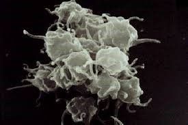
Source of images: http://www.ouhsc.edu/platelets/platelets/Platelet%20Pics/Platelets%201.gif
http://images.medicinenet.com/images/featured/detail_blood_clot.jpg
The heart: The heart pumps blood through veins and arteries around the body. Arteries carry blood from the heart. Veins carry it towards the heart. When you breathe in and breathe out your heart contracts and expands. The artery is big close to the heart because there is lots of pushing force. If the artery is small then it will burst. Gradually the blood vessels become smaller because there is less blood and hardly any force. Then when the heart expands a vacuum is created which pulls the blood back to the heart. Slowly the veins become bigger because the more close you go the more force there is. Also more blood joins in.
The heart generates high pressure to pump the blood out of the heart and low pressure to get it back. The heart beats 60 times per minute.
The outer layer of the heart (fibrous pericardium) is made of dense connective tissue. It helps protect the heart and anchors it. The second set of layers is called the serous pericardium. It contains: The parietal pericardium. Then is serous fluid which is a fluid. It lets the heart beat and stops the creation of friction. Then comes the visceral pericardium. Then comes the myocardium which is mainly cardiac muscle tissue that does the work of contracting. The next is endocardium. This is a thin white layer of squamous epithelial tissue( layer of skin).
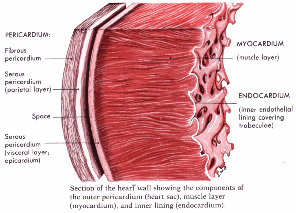
Source of image: http://stevegallik.org/sites/histologyolm.stevegallik.org/images/heartwall.gif
The heart is divided into 2 parts by a thin inner partition called the septum. This makes 4 chambers. Two superior atria(low pressure areas) and two inferior ventricles(high pressure areas). Each chamber has a valve that lets blood through not back. It has 4 valves. The mitral(bicuspid valve)( it has 2 flaps) and tricuspid valve(it has 3 flaps) and the aortic and pulmonary semilunar valves. The right ventricle pumps blood through the pulmonary semilunar valve into the pulmonary trunk (a big vessel that splits to make the right and left pulmonary arteries).
The blood then goes straight to the lungs. In there it goes in the thin walled capillaries. It picks up oxygen and leaves carbon dioxide. Then goes back to the heart to 4 pulmonary veins. It moves to the left atrium. Then this contracts so the blood passes down to the mitral valve into the left ventricle. This whole thing is called the pulmonary circulation loop. The left ventricle flexes creating pressure. The blood doesn’t go back into the left atrium because the mitral valve shuts. Then the blood has to go through the aortic semilunar valve into the body’s largest artery. Its called the aorta. From here it gets sent to the rest of the body.
After the loop the blood comes back through the big superior and inferior vena cava veins into the right atrium. When this contracts blood passes through the tricuspid valve into the relaxed right ventricle. Then it starts over. When you hear someone’s heart beating you hear a lub dub lub dub. The lub is made by the mitral and tricuspid valves closing. This is when the blood gets pumped out of the heart(systole). The dub is made by the aortic and pulmonary semilunar valves closing. This is when the blood comes from the atria (diastole). Blood is always red. Deoxygenated blood is just dark red.
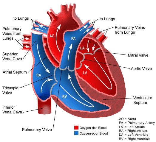
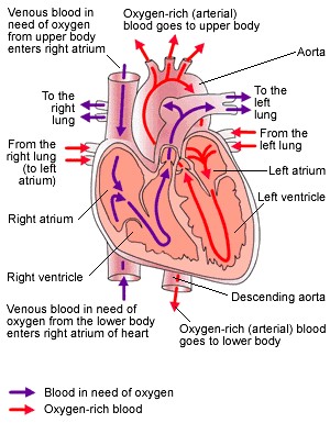
Source of images: http://projects.ecfs.org/prepole/PHYSIOLOGY%2013/Heart%20and%20Circulation%20System/Pictures/Heart%20Diagram.jpg
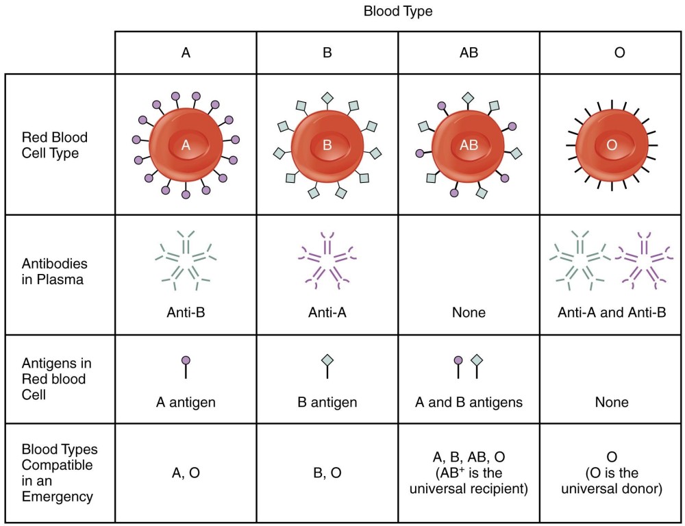
Blood type: All the cells in your body have antigens. They are protein markers that are like name tags. There are 2 main types of antigens that are on your RBC. The 1st is agglutinogens. The 2 kinds of agglutinogens are named A and B. The 2nd is called the rhesus or the Rh system. The Rh system are a collection of 45 different antigens. These all produced as one group. You either have all of them or none of them. People who have the collection are Rh positive. And people who don’t have are Rh negative.
There are 8 blood types. A, B, AB, O and a positive and negative for each type. Blood type is heredity. For example If one parent has B and the other has a the baby’s blood type would be either A or B or AB. In order for someone to have an O blood type both parents have to have O. AB blood types have no antibodies. They can accept all of the types. Type O have antibodies for A and B. They can accept only O type. Whereas Rh positive can accept both but Rh negative can only accept negative.
Antigens are proteins or sugar. When you get a blood transfusion then when the blood enters our body then the immune system dispatches antibodies to check if they are foreign or not. They attach themselves to the new cells that have entered our body. If they are foreign then the antibody terminates them.
When an antibody attaches to a cell it connects with the cells antigens. If it recognises the configuration then it leaves it. O blood types don’t have antigen on their cells so there blood can be given to anyone because the other person’s antigens will not be able to attach themselves to the given O type blood cells. So it won’t be able to recognise it. These are the universal donors. AB are universal receptance.
When you get the wrong type of blood it could cause an acute haemolytic transfusion reaction. When the immune system attacks the foreign cells they are killed rapidly. This could result in a pile up and this could cause a vein to get blocked. B cells are wbc that make antigens.
Antibodies are also called immunoglobulins. The genes which are in your cells who have a nucleus contain which blood type you are. There are 5 type of antibodies. IgG IgM IgA IgD IgE. They are produced by plasma cells.
T cells destroy antigens. T cells also help signal other cells. Also antibodies activate a group of proteins called complements to assist them in killing. When complements come then the foreign substances get coated with antibodies. The arms of the antibodies are pointy so when they make contact with a foreign cell it makes a hole so it bursts.
Source: http://philschatz.com/anatomy-book/resources/1913_ABO_Blood_Groups.jpg
Blood
| Plasma | RBC | WBC | Platelets |
| regulates fluids, helps to remove waste, controls electrolyte balance. | transports oxygen, brings back co2. | attack bacteria viruses and germs. they kill pathogens. | they clog a rupture in the vessel. |
| contains: 90% water. 10% other things like enzymes and proteins. | Contains hemoglobin | ||
| pale yellow | red | white | white |
| no nucleus | nucleus | ||
| liquid | cell | cell | fragment of cell |
Bibliography
1: https://en.wikipedia.org/wiki/Red_blood_cell
2: https://www.youtube.com/watch?v=X9ZZ6tcxArI
3: https://en.wikipedia.org/wiki/White_blood_cell
4: http://www.urmc.rochester.edu › Health Encyclopedia
5: www.healthline.com/health/wbc-count
6: https://en.wikipedia.org/wiki/Blood_type
7: http://www.nhs.uk/conditions/blood-groups/Pages/Introduction.aspx
